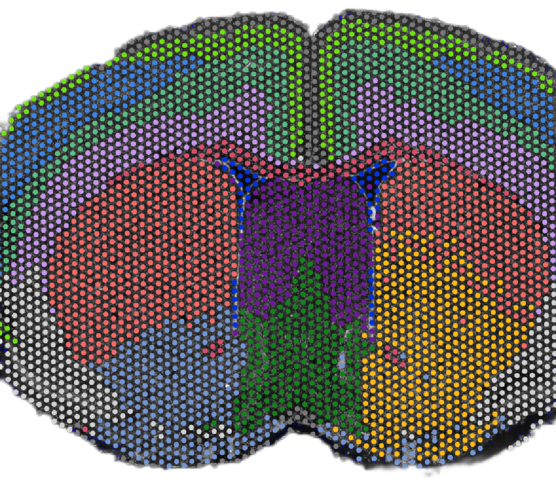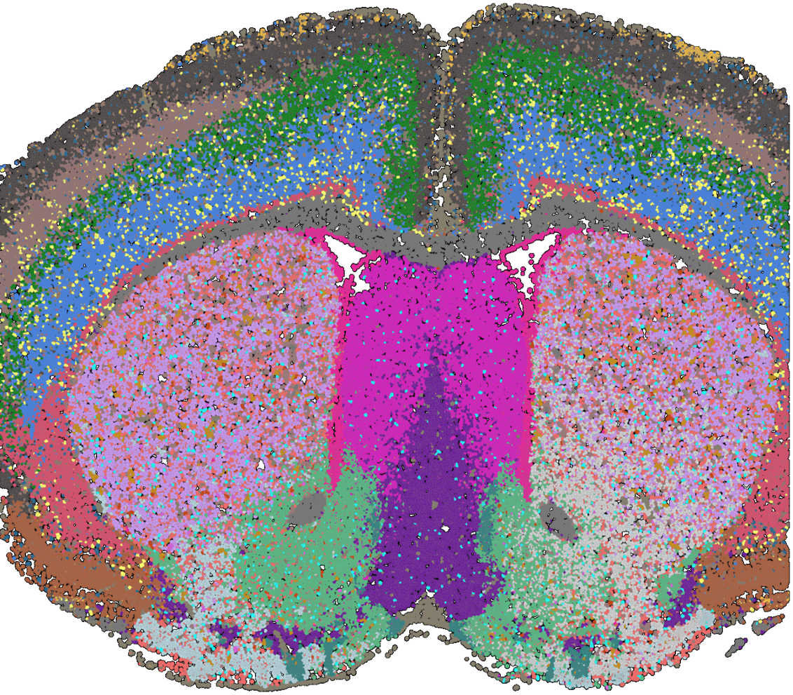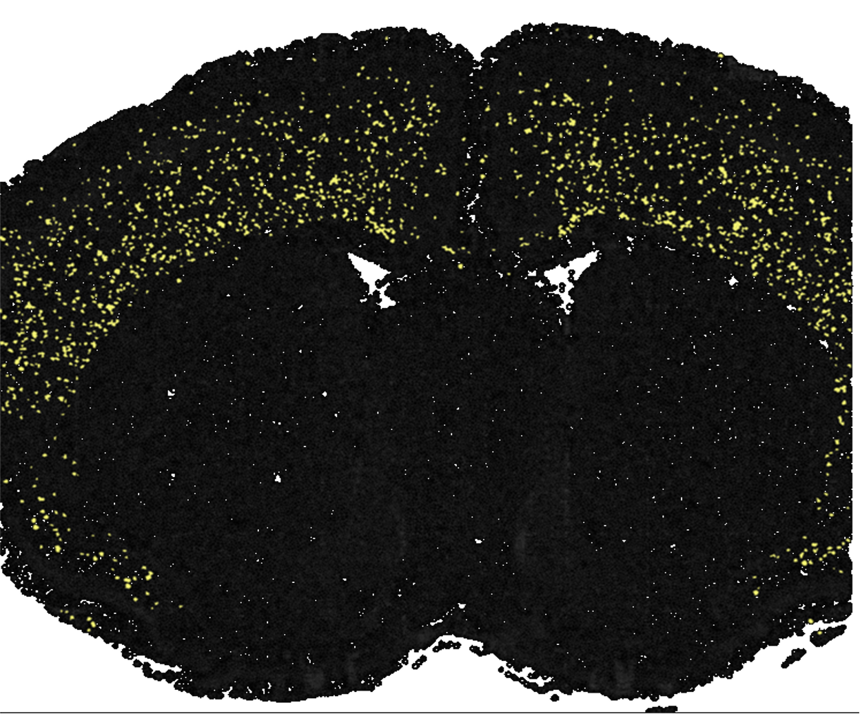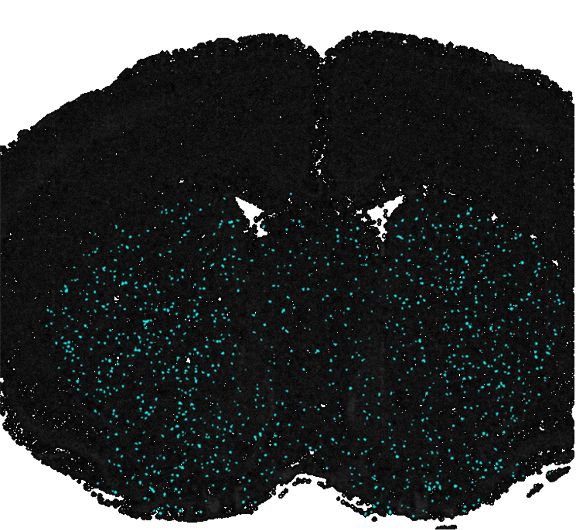

Single-Cell Level Spatial Transcriptomics Platform: BMKMANU S3000
BMKMANU S3000 enables whole transcriptome spatial analysis at single-cell resolution, built upon our proprietary microwell-array architecture, with barcoded microspheres loaded into each well. A total of 4,144,770 spots are arranged across a 6.8 × 6.8 mm capture area, with each bead conjugated to ~105 oligos. This high-density bead and oligo configuration achieves subcellular resolution while maintaining high capture efficiency. Integrated with high-definition H&E and fluorescence imaging, our proprietary image stitching and calibration technology supports precise cell segmentation and clustering. BMKMANU S3000 is compatible with mainstream second- and third-generation sequencing platforms, as well as multi-omics workflows including BCR-seq & TCR-seq and ATAC-seq.
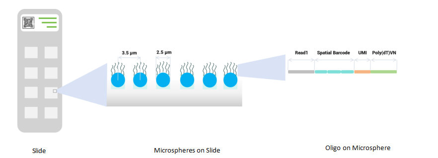
- Subcellular resolution (3.5 μm)
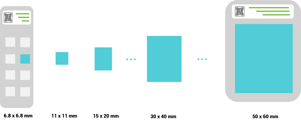
- Multiple capture area sizes are available to suit diverse applications
- Excellent RNA capture performance with ~88% effective capture area
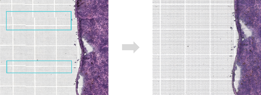
- Reduced lateral RNA diffusion due to bump slide surface
- Precise cell segmentation based on high-definition H&E and fluorescence imaging, as well as proprietary image stitching and calibration technology
- Compatible with second- and third-generation sequencing platforms: Illumina, MGI, GeneMind, ONT, PacBio, etc.
- Compatible with multiomics workflows, including BCR-seq/TCR-seq and ATAC-seq
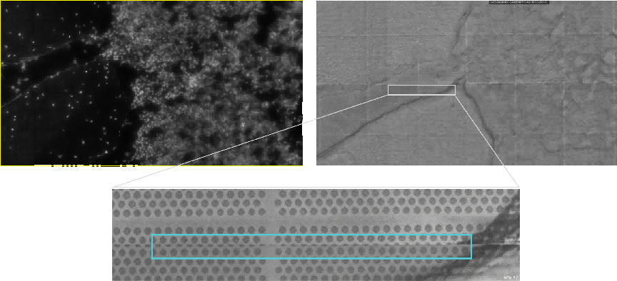
- Cell segmentation for animal cells

fluorescence image
based on H&E image
- Cell segmentation for plant cells

toluidine blue staining
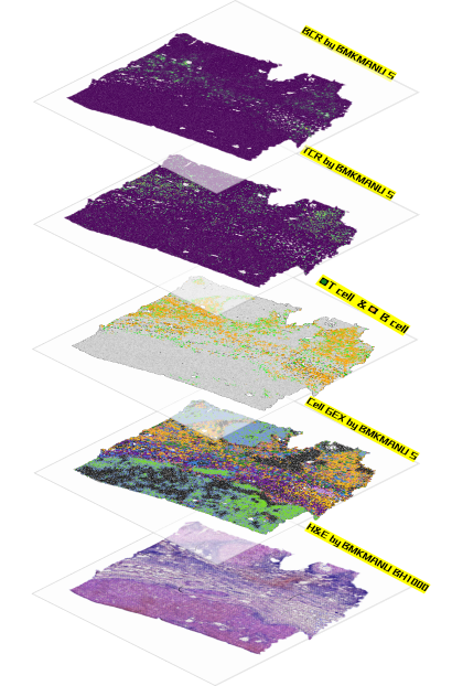
Case Study of S3000 Spatial Transcriptomics Platform
Number of Reads: 1,647,163,344
Sequencing Saturation: 68.67%
Median Genes per Cell: 1,999
Median Genes (30μm): 3,793
Median Genes (18μm): 2,026
Median Genes (6μm): 510