

Introduction of BH1000 Spatial Omics Imaging System
BMKMANU BH1000 Spatial Omics Imaging System is compatible with BMKMANU S-series spatial omics platform and supports imaging through the whole experimental workflow. Capable of fast whole-slide scanning for multiple specimens from diverse species, it delivers high-precision, high-resolution bright-field and fluorescence images. Integrated with BMKMANU’s proprietary cell-segmentation algorithm, it achieves accurate spatial single-cell segmentation.
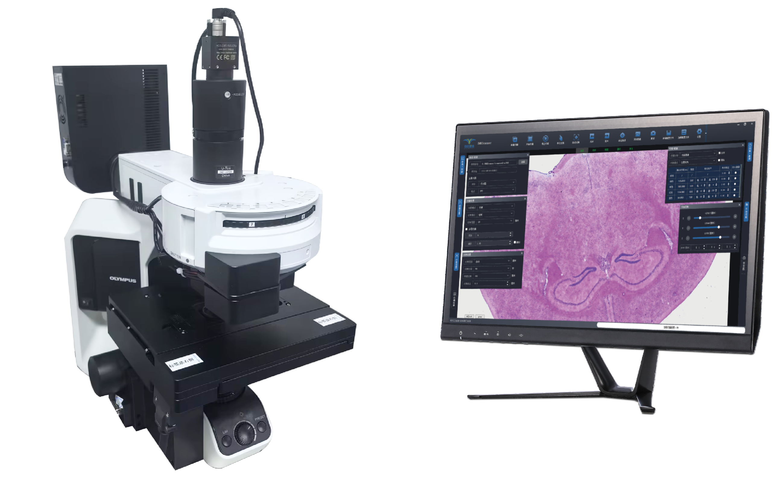
Features of BH1000 Spatial Omics Imaging System
Easy Operation | User-friendly Interface
Ultra-fast Scanning | Large Scanning Area
Compatible with Multi-layer Fusion Scanning
Multi-region Scanning
Compatible with BMKMANU S-series Spatial Omics Platform
It supports error-free high-resolution H&E & fluorescence imaging for S-series spatial omics slides to achieve subcellular-resolution spatial omics data.
Error-free Image Stitching for Precise Cell Segmentation
The embedded error-free image stitching calibration algorithms based on the microsphere-array architecture of S-series slides, combined with cell-segmentation algorithms, lead to precise cell segmentation.
Scanning Cases of BH1000 Spatial Omics Imaging System

Spatial Omics Clustering Results Using BH1000 Spatial Omics Imaging System

Specifications of BH1000 Spatial Omics Imaging System
| Parameters | Descriptions |
|---|---|
| Scanning Area | Preview of scanning area available; 30 mm×60 mm; Maximum 50 mm×60 mm (optional) |
| Resolution | 0.28 μm/pixel (with 20× objective) |
| Scanning Speed | Scanning time ≤55 s for an 8 mm×8 mm scanning area under 20× objective |
| Scanning Modes | Single-layer, multi-layer, multi-layer fusion |
| Objective Lenses | Standard: 10× objective (NA 0.25); 20× plan-apochromatic objective (NA 0.8). Optional: 40× objective (NA 0.95) |
| Light Source System | Bright-field: high-stability LED light source. Fluorescence: LED stroboscopic light source to effectively prevent photobleaching |
| Fluorescence Module | Multi-channel fluorescence module, max. 6 channels. Each channel equipped with independent sensor with motorized switching. Standard channels: Bright-field, DAPI, FITC, and CY3 |
| Spatial Omics Compatibility | Compatible with spatial-omics slide, which enables error-free H&E and fluorescence imaging, leading to precise cell segmentation for spatial transcriptomics clustering. Compatible with multi-omics profiling: Spatial RNA-seq & spatial BCR-seq & TCR-seq & spatial ATAC |