

New Release | BMKManu DG1000 Single-Cell RNA Sequencing Arrives
BSTViewer v3.0 Installation Tutorial
2022-06-08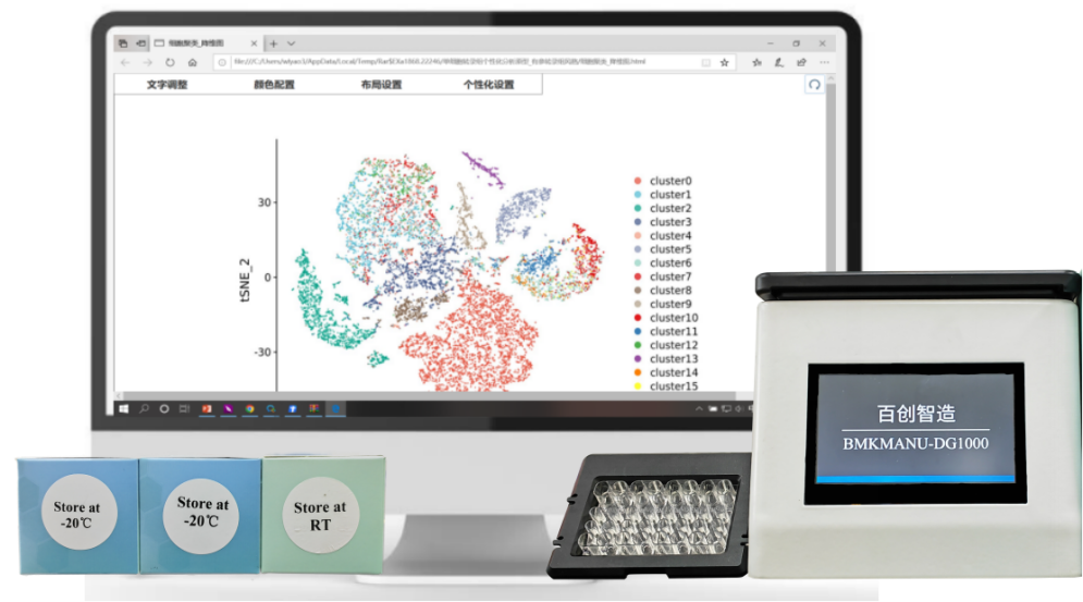
BMKManu DG1000 vs. Mainstream Overseas Single-Cell Platforms
2022-06-21
The heterogeneity of gene expression and function among individual cells determines cell fate during development and is closely tied to the mechanisms of disease. Single-cell sequencing makes it possible to sequence the transcriptome, and even the genome, at the level of individual cells, bringing gene sequencing down to the single-cell scale, improving accuracy and opening up entirely new possibilities for biotechnology R&D and application.
After years of accumulated technological expertise, Biomarker released its innovatively developed single-cell microdroplet system — the BMKManu DG1000. As Prof. He Shunping of the Institute of Hydrobiology, Chinese Academy of Sciences, noted at the BMKManu DG1000 launch event, single-cell sequencing is an innovative gene sequencing technology, but its relatively high library construction cost has been an obstacle preventing it from reaching ordinary laboratories. The BMKManu DG1000 largely solves the problem of high single-cell sequencing costs, which will greatly increase research output and allow more researchers to benefit from single-cell technology.
[su_heading size=”16″]Overview of BMKManu DG1000 single-cell RNA sequencing technology[/su_heading]
BMKManu DG1000 uses microfluidics, oil droplet encapsulation and barcode labeling to achieve high-throughput cell capture. It completes the water-in-oil process within a short time to form an emulsion, and rapidly supports flexible processing of 1–8 samples to build single-cell sequencing libraries. It can be applied to cell atlas construction, tumor heterogeneity, neuroscience, immune infection, stress response, biological evolution and other fields, and will become a powerful tool in clinical medicine and basic research.

[su_heading size=”16″]BMKManu DG1000 end-to-end single-cell service[/su_heading]
Biomarker provides an end-to-end single-cell solution covering study design, single-cell suspension preparation, single-cell sorting, library construction and sequencing, bioinformatics analysis, customized analysis and bioinformatics training, helping researchers conduct single-cell transcriptomics studies with high throughput and low cost.

Figure 1. Schematic of the BMKManu DG1000 single-cell RNA sequencing workflow
[su_heading size=”16″]Technical advantages[/su_heading]

[su_heading size=”16″]Sample requirements[/su_heading]
- Sample types
1) Tissue samples: after harvesting fresh tissue (about 200 mg) from a living organism, remove surrounding fat, connective tissue or necrotic tissue, rinse thoroughly with pre-cooled PBS or saline, and transfer it directly, or in tissue preservation solution, to the dissociation laboratory as quickly as possible for single-cell suspension preparation;
2) Blood samples: collect 2–3 mL of fresh blood and store it immediately in an EDTA anticoagulant tube (purple top), then transfer or ship it to the Biomarker laboratory at 4°C (generally within 12 h) and isolate PBMCs before loading; alternatively, isolate PBMCs by density gradient centrifugation, add cell freezing medium and freeze in a stepwise manner, then ship on dry ice;
3) Fresh frozen samples (single-nucleus RNA sequencing): rinse fresh tissue clean, place it in a cryotube, snap-freeze in liquid nitrogen, store at -80°C, and finally ship on dry ice;
- Single-cell suspension requirements
More than 5×10⁵ cells, viability greater than 85%, cell diameter less than 60 μm, aggregation rate <15%, few debris, and no reverse transcription inhibitors or non-cellular nucleic acid molecules.
[su_heading size=”16″]Application scenarios[/su_heading]
The application of single-cell sequencing technology links cell types to cell fate, state and function, advancing research fields such as developmental biology, oncology, neuroscience, immune infection and stress response.
- Atlas construction
By constructing single-cell atlases of different species, tissues, diseases and cell subtypes, gene expression patterns across all cell types can be determined and specific marker genes identified. The Fly Cell Atlas: a single-nucleus transcriptomic atlas of the adult fruit fly[1], published in Science in 2022, performed single-cell transcriptomic analysis of 15 tissue types, 63 samples and a total of 580,000 nuclei from Drosophila, building a comprehensive Drosophila cell atlas;
- Growth and development
To study developmental and differentiation processes such as seed germination, embryonic development, organ formation, tissue regeneration and tumor/disease progression, and to reveal the developmental differentiation trajectories of different cell types. Molecular architecture of the developing mouse brain[2], published in Nature in 2021, sampled the embryonic mouse brain daily from day 7 to day 18, obtaining 215 embryos, 93 samples and a total of 292,495 cells, and constructed a mouse brain atlas spanning the developmental process;
- Disease molecular mechanisms
To explore changes in cell composition and gene expression in tumor and disease environments, study immunological questions such as immune recognition and response, and reveal the functions of different cell types and the molecular mechanisms of disease onset in tumor and disease contexts. A pan-cancer single-cell transcriptional atlas of tumor infiltrating myeloid cells[3], published in Cell in 2021, performed single-cell transcriptomic analysis of tumors, adjacent normal tissues, peripheral blood and lymph nodes from 194 patients — 338 samples and a total of 138,161 myeloid cells — and explored the composition of tumor-infiltrating myeloid cells, a key regulatory factor in tumors, and its correlation with clinical factors.

Figure 2. Construction of large-scale single-cell atlases [1,2,3]
As single-cell technology becomes more widely used, requirements for publishing single-cell papers are also rising. Building fine-grained cell atlases of hundreds of thousands of cells or more by stacking large-scale samples, in order to mine rare cell subpopulations and reveal entirely new molecular regulatory mechanisms, has become the standard approach for publishing CNS-level papers.
The arrival of the BMKManu DG1000 lowers the barrier to constructing large-scale cell atlases, allowing more researchers to benefit from cutting-edge technology
References:
- Li H, et al. “Fly Cell Atlas: a single-nucleus transcriptomic atlas of the adult fruit fly.” Science (New York, N.Y.) vol. 375, 6584 (2022): eabk2432. doi:10.1126/science.abk2432
- La Manno G, et al. “Molecular architecture of the developing mouse brain.” Nature vol. 596, 7870 (2021): 92-96. doi:10.1038/s41586-021-03775-x
- Cheng S, et al. “A pan-cancer single-cell transcriptional atlas of tumor infiltrating myeloid cells.” Cell vol. 184, 3 (2021): 792-809.e23. doi:10.1016/j.cell.2021.01.010
Related posts


Warning: Trying to access array offset on value of type bool in /www/wwwroot/10.3.128.39/wp-content/themes/betheme/functions/theme-functions.php on line 1467
Warning: Trying to access array offset on value of type bool in /www/wwwroot/10.3.128.39/wp-content/themes/betheme/functions/theme-functions.php on line 1467